Iron deficiency is far more than fatigue and low haemoglobin: because iron underpins oxygen delivery, enzyme activity, and connective-tissue synthesis, shortages frequently show up first on the skin, hair, and nails. Patients commonly report a spectrum of visible changes — subtle pallor, persistent dryness or itch, painful mouth cracks and a smooth tongue, brittle or spoon-shaped nails, diffuse hair thinning, and slower wound closure. These signs may precede laboratory confirmation and, when recognised, prompt targeted testing (haemoglobin, ferritin, transferrin saturation) and treatment that often reverses dermatologic symptoms. This article explains the biological basis for these findings, describes how clinicians interpret tests and choose therapy, and offers practical, evidence-based guidance for patients and clinicians aiming to restore both iron stores and skin health.
Why iron matters to skin, hair and nails
Iron is a catalytic cofactor in key biochemical reactions required for epidermal cell proliferation, collagen synthesis, and keratin formation. Adequate iron supports mitochondrial energy production and oxygen delivery via hemoglobin; both are essential in tissues with rapid cell turnover, like the epidermis and hair follicles. When iron is lacking, collagen cross-linking and extracellular matrix remodeling are impaired, keratin structure weakens, and tissue oxygenation drops — a combination that produces drier, thinner skin, fragile nails, and hair growth disturbances. These mechanistic links explain why cutaneous signs are common and why they often improve once iron is repleted.
Iron deficiency symptoms on skin

- Pallor (pale skin and mucous membranes)
The most direct dermatologic consequence of iron deficiency is pallor. Reduced hemoglobin lowers the red coloration in capillary blood and makes skin and mucous membranes (conjunctiva, inner eyelids, palmar creases) appear lighter. Pallor is a sensitive but non-specific clinical sign—its visibility varies with baseline skin tone—so clinicians inspect conjunctivae and nail beds when pallor is suspected.
- Dryness, pruritus and impaired barrier function
Chronic iron shortage diminishes keratinocyte proliferation and weakens barrier repair, producing xerosis (dry skin) and pruritus. Patients may describe persistent flaking, winter-worse itch, or fissures in high-stress areas. Several observational studies and clinical reviews link low iron/ferritin states to these symptoms; importantly, they often improve with iron repletion.
- Angular cheilitis and glossitis (mouth findings)
Iron deficiency is a well-documented cause of angular cheilitis (painful cracks at the lip corners) and atrophic glossitis (a smooth, sore tongue). These mucosal changes reflect epithelial atrophy and impaired mucosal repair and are frequently reversible after iron correction. Because similar mouth findings occur with B-vitamin deficiencies and candidiasis, clinicians often test multiple nutrient markers.
- Koilonychia (spoon-shaped nails) and brittle nails
Chronically low iron can lead to thinning and deformation of the nail plate; koilonychia — concave, spoon-shaped nails — is a classic sign of long-standing deficiency. While not universal, its presence strongly raises suspicion for chronic iron loss or malabsorption.
- Hair thinning and telogen effluvium
Iron deficiency is associated with diffuse hair shedding (telogen effluvium) and worsened hair quality. Research shows lower ferritin levels in many patients with nonscarring alopecia; clinicians commonly include ferritin in hair-loss workups. Hair recovery after iron repletion can take months because follicles progress slowly through growth cycles.
- Slow wound healing and increased fragility
Because iron contributes to collagen deposition and immune competence during wound repair, deficiency can slow re-epithelialization and remodeling, making wounds take longer to close and increasing infection risk. Nutrition-and-wound-healing reviews document iron’s role in multiple healing phases.
Preventive measures for iron deficiency
- Eat
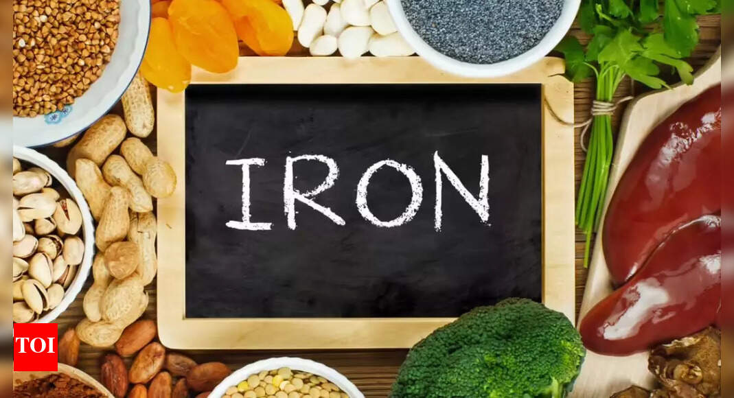
iron-rich foods regularly – Include heme iron (red meat, poultry, fish) and non-heme iron (beans, lentils, spinach, fortified cereals). - Boost absorption with Vitamin C – Pair meals with citrus fruits, tomatoes, bell peppers, or strawberries to enhance iron absorption.
- Limit iron inhibitors – Avoid tea, coffee, or calcium-rich foods immediately with iron-rich meals as they block absorption.
- Choose fortified foods – Opt for iron-fortified cereals, flours, and plant-based milk for additional intake.
- Monitor blood loss – Seek medical advice for heavy periods, frequent nosebleeds, or gastrointestinal bleeding to prevent chronic iron loss.
- Routine screening – Pregnant women, infants, vegetarians, and older adults should have regular hemoglobin and ferritin checks.
- Use supplements when prescribed – Take iron tablets or prenatal vitamins as directed by a doctor—self-supplementation can be harmful.
Disclaimer: This article is for informational purposes only and is not a substitute for professional medical advice. Consult a healthcare provider for diagnosis, treatment, or before starting supplements or dietary changes.Also Read | This everyday nut could be the secret to lower cholesterol, heart disease prevention, and longevity


